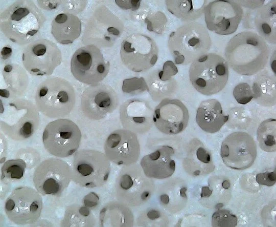
Spherical porous architecture of interconnected pores, within each pore, provides a truly connected structure.
A better bioactive approach to stabilization:
Pores within Pores
A Scaffold for bone in-growth and long-term fixation: Each fugitive material is manufactured in-house and perfectly spherical. We eliminate all non-spherical fugitive materials, leaving only spherical materials allowing for interconnectivity. Pore Matrix interconnected spherical architecture increased bone ingrowth in sheep, showing 1 mm of bone growth at 4 weeks and up to 3 mm of bone growth at 12 weeks using the non-biologic version. Future studies are being conducted on several bioactive porous versions with our partners.
Porous PEEK cancellous
Non-Bio Active Porous PEEK sample
Histology confirmed the findings from micro-computed tomography. The Initial stability for early fixation at 4 weeks with 1mm ingrowth has limited micro motion. Micro computed tomography was a practical endpoint in the current study, especially at 12 weeks, where osseointegration into the porous PEEK was easy to observe. New bone was found at the host margins in the cortical sites in the porous PEEK samples at 12 weeks.
Consolidated into Cortical Bone
The strength required for weight bearing. Peak load and shear stress at 4 and 12 weeks. Note, the Porous PEEK samples consolidated during testing and were not able to be pushed out completely. The properties of Porous PEEK improved with time.

Let the Data talk
“ I firmly believe as the spine industry continues to evolve, the need for titanium is no longer the answer. This is. It remodels and is strong! My doctor used an HA PEEK cervical interbody and within one year it looks like bone… all you see is markers. This will only work more efficiently and effectively through real integration.”
Industry Professional
12-week implantation cancellous
The Porous PEEK sample in cancellous bone at 12 weeks demonstrated areas of integration into the porous material integrated at this time point (arrows). Non-Bioactive Porous PEEK implants exemplified areas of bone integration that penetrated a few millimeters into the implants by 12 weeks in cortical and cancellous sites.
Cortical bone ingrowth 12 weeks
Porous PEEK sample in cortical bone at 12 weeks demonstrating areas of integration into the porous material at the cortical host bone margins (arrows). Non Bioactive Porous PEEK implants in the current study supported osseointegration. Osseointegration improved the mechanical performance of the porous PEEK in push out testing.


